Equine Veterinary Journal Early View November 2015
By Heather Ferguson
An anatomical study of the dorsal and ventral nasal conchal bullae in normal horses: Computed tomographic anatomical and morphometric findings .
Liuti, R. Reardon, S. Smith and P.M. Dixon
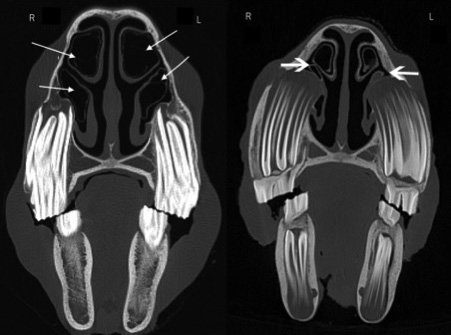
Involvement of dorsal and ventral nasal conchal bullae can be clinically important in sinusitis. This study aimed to describe the computed tomographic anatomy of these structures: 90 equine heads were obtained from abattoirs and their age estimated from dentition. Computed tomography images were acquired and examined by diplomats. Specimens with dental or sinonasal disease (confirmed by skull sectioning and gross examination) were excluded from the study, leaving 60 specimens.
Imaging software (OsiriX) was used to create multi-dimensional reconstructions and measure height, length and width of dorsal and ventral bullae allowing the volume of each bulla to be calculated. Height, length and width of each head also provided a volume for each skull.
The dorsal conchal bullae were found to be greater in volume (average 24 cm3) than ventral conchal bullae (average 15cm3) and these were related to head size. In both dorsal and ventral bullae, there were significant differences in the volumes between different age groups, with those in the youngest group (0–5 years) having significantly smaller volumes than those in the oldest group (over 16 years). In the case of the ventral conchal bullae, this could be explained by the larger size of cheek teeth alveoli protruding into the nasal cavity in younger horses.
In the majority of horses (81.3%) the rostral limit of the dorsal conchal bulla was parallel to Triadan 07s. The caudal limit was parallel to Triadan 10s in 61% and the 09s in 30.5%. The rostral limit of the ventral conchal bulla was parallel to Triadan 07s in 78% and the caudal limit parallel to the 09s in 64.5% and the 10s in 32%. This shows that in some horses the caudal limits of the ventral conchal bulla can overlap with the rostral extent of the rostral maxillary sinus.
Bottom line:
This study provides information on the anatomical features of dorsal and ventral conchal bullae and their relationship with adjacent structures and will help in identifying which structures are involved in sinus disease.
--Ends--