Subclinical ultrasonographic abnormalities of the suspensory ligament branches in National Hunt racehorses
A.J. Fairburn, E. Busschers and A.R.S. Barr
This cross-sectional study conducted on 62 horses at a single National Hunt training yard aimed to investigate the prevalence of subclinical ultrasonographic abnormalities of the suspensory ligament branches (SLBs). It also aimed to establish cross-sectional areas of suspensory ligament branches in the National Hunt population. Horses with a history of suspensory ligament injury or any abnormality detected on palpation during veterinary examination were excluded from the study.
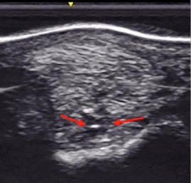
A standardised set of 10 images of the SLB were obtained for the forelimbs of each horse, anonymised images were assessed by three clinicians. A previously reported grading system was used to score each site from 0 to 3, with the highest scores from each site being used to formulate a ‘branch grade’. The cross-sectional area (CSA) at each site was also measured and a mean area calculated.
There was good inter-observer agreement: 58 SLBs were graded as Grade 0, 163 as Grade 1 and 27 as Grade 2. Of the 30% of the population with a Grade 2 score, all had abnormalities at the insertion and almost all these abnormalities were palmar-abaxial on the transverse images and abaxial/central-abaxial on the longitudinal images. There were significantly more Grade 2 lesions in the medial SLBs compared to lateral SLBs, possibly related to asymmetrical loading of the medial branches.
The mean CSA of medial and lateral branches was between 1.3 and 1.4 cm2 but CSA was an insensitive indicator of subclinical injury. CSA was significantly larger in the medial SLBs than lateral SLBs and the authors advised comparing CSA with the corresponding branch in the contralateral limb to identify unilateral enlargement. All abnormalities were classed as sub-clinical as no horse developed SLB injury in the season following scanning.
Bottom line:
A third of this National Hunt racehorse population had Grade 2 ultrasonographic abnormalities of the SLB. Medial SLBs were more prone to Grade 2 lesions than lateral SLBs. None of these ultrasonographic abnormalities were associated with clinical injury. The mean cross-sectional area of the medial SLB was larger than the lateral SLB, hence comparison with the contralateral limb is advised.
--Ends--